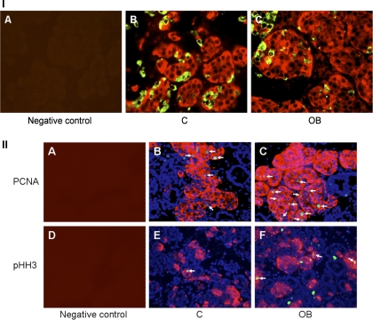
Fig. 5.
Immunofluorescent staining of the fetal pancreas. I: pancreatic sections stained with either no first antibody as a negative control (A), or insulin (red) and glucagon (green) in both control (B) and obese (C) groups. II: cell proliferation test using immunofluorescent staining of PCNA and pHH3. Pancreatic sections stained with no first antibody as a negative control (A and D). Insulin (red) and PCNA (green) positive cells stained for counting in both control (B) and OB (C) groups. Insulin (red) and pHH3 (green) cells stained for counting in both control (E) and OB (F) groups. DAPI (blue) indicates general nuclear staining. Arrows indicate PCNA (B and C) and pHH3 (E and F) positive β-cells.

