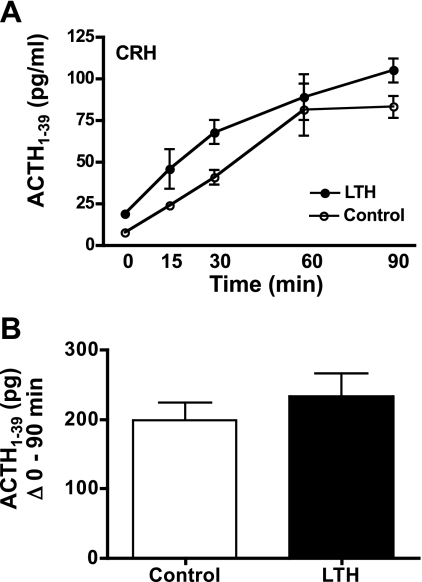
Fig. 3.
A: plasma concentrations of ACTH1-39 in LTH and control fetal sheep during infusion of CRH. CRH infusion was initiated immediately after the preinfusion sample (n = 4 per group; means ± SE). Concentrations of ACTH1-39 were significantly elevated (P < 0.05) in response to CRH in both groups. ACTH1-39 was significantly higher (P < 0.05) in LTH compared with control fetuses prior to and during CRH infusion. B: sum of fetal plasma ACTH1-39 for LTH and control fetuses from 0 (pre-CRH) through 90 min of CRH infusion. There were no differences in total ACTH1-39 released between groups.

