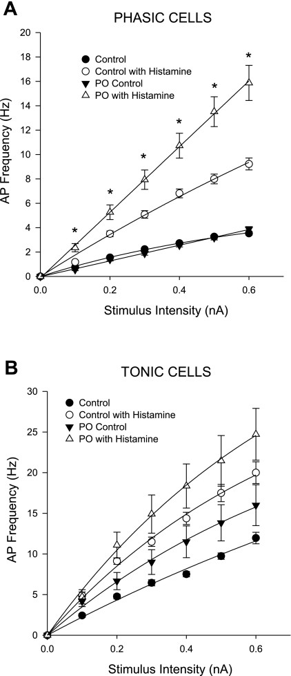
Fig. 3.
Up-regulation of the histamine-induced increase in neuronal excitability is observed in phasic intrinsic cardiac neurons with pressure overload. The frequency of evoked APs (means ± SE) as a function of increasing stimulus intensity (0.1–0.6 nA, 500-ms duration) was determined in intracardiac neurons derived from control and PO models prior to and following histamine application. Data from control (untreated) and sham surgical animals were not significantly different and were pooled. Neurons were categorized as either phasic (A, top) or tonic (B, bottom), based on their firing responses to a prolonged stimulus. There were no significant differences in the frequency of evoked APs in either phasic or tonic cells from PO vs. control tissues prior to histamine application. Histamine application induces a significant increase in AP frequency in both phasic and tonic neurons. However, the histamine-induced increase was significantly greater in phasic neurons from PO tissues compared with the histamine-induced responses in phasic neurons from control preparations. Conversely, there was no significant difference in the histamine-induced responses in the tonic neurons from PO and control preparations. The frequency vs. stimulus curves for the phasic neurons from PO preparations were best fit with a linear equation (R2 > 0.99 for both), while the remaining curves were best with a single exponential curve (R2 > 0.99). Statistical significance was evaluated at each stimulus intensity for a given treatment (i.e., control with histamine vs. PO with histamine) by t-test, *P < 0.05.

