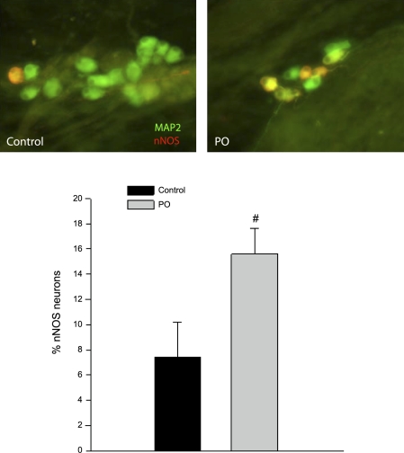
Fig. 6.
PO induces an increase in neuronal nitric oxide synthase (nNOS)-expressing neurons in the cardiac plexus. Whole mounts of the cardiac plexus from control and PO animals were fixed and stained for immunohistochemical analysis. The percentage of nNOS neurons was determined by antibody labeling for nNOS (rabbit anti-nNOS 1:500 and donkey anti-rabbit-Cy3 1:500, red) and MAP2 (mouse anti-MAP2 1:500 and donkey anti-mouse FITC 1:500, green). Neurons with colocalization appear yellow or orange in color. Top: an example of nNOS staining in a control ganglia vs. a ganglia from a PO animal is shown. The percentage of neurons that stained positively for both MAP2 and nNOS was calculated in six preparations from control and PO tissues, and the means ± SD are shown in the bottom panel (black bar, controls; gray bar PO) with a significant increase (#P < 0.001) in the percentage of nNOS cells observed in the PO preparations.

