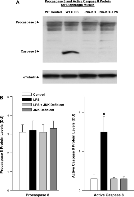
Fig. 6.
Caspase 8 protein levels in JNK-deficient animals. A: Western blots stained for procaspase 8 and cleaved. Active caspase 8 protein (top) are representative diaphragm samples. α-Tubulin protein levels for these same samples (bottom) served as a loading control. LPS administration elicited a large increase in active cleaved caspase 8 protein compared with the control. This increase was not seen in JNK-deficient animals given LPS. The sample from the JNK-deficient animal given saline had caspase levels similar to control. WT, wild type; KO, knockout. B: mean data examining the effect of LPS on diaphragm caspase activation in JNK-deficient animals. Densitometry was used to quantitate procaspase 8 and active caspase 8 protein levels for diaphragm samples. Procaspase 8 levels were similar for the 4 experimental groups (left). In contrast, diaphragm-active caspase 8 protein levels increased dramatically for LPS-treated animals (P < 0.001). Administration of LPS to JNK-deficient animals did not cause an increase in active caspase 8 protein (*P < 0.01 for comparison between LPS and LPS + SP600125 groups).

