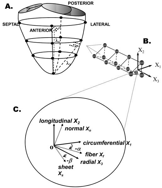
FIGURE 1.
A. Locations of LV epicardial markers (large filled circles), LV lateral equatorial and anterior basal transmural beadsets (small filled circles). B. Transmural tissue blocks for histological measurements were excised from each heart at the anterior basal and lateral equatorial regions immediately below the transmural beadsets, with the edges cut parallel to X1, X2, and X3 , the circumferential, longitudinal and radial cardiac axes, respectively. C. Transmural fiber angles (α) were measured from sections cut parallel to X1-X2 plane. At a given wall depth, measured fiber angles (α) and sheet angles (β) were used to define local fiber-sheet coordinates with basis vectors of fiber (Xf) axis, sheet axis perpendicular to Xf within sheet plane (Xs), and axis normal to the sheet plane (Xn). These same measurements were obtained from the anterior wall tissue block.

