
Figure 1.
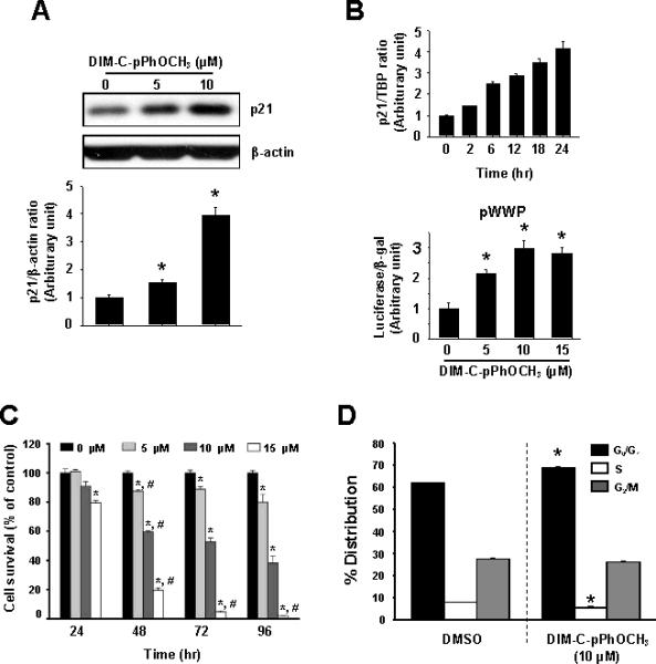
DIM-C-pPhOCH3 induces p21 and decreases Panc1 cell proliferation. Induction of p21 protein (A), mRNA levels, and promoter activity (B). Panc1 cells were treated with 0, 5 and 10 μM DIM-C-pPhOCH3 for 24 hr (A) or 10 μM DIM-C-pPhOCH3 for different times (B), and cellular extracts were analyzed by Western blots or real time PCR as described in the Materials and Methods. For promoter activity, cells were transfected with pWWP, treated with DIM-C-pPhOCH3 and luciferase activity determined as described in the Materials and Methods. (C) Panc1 cell survival. Panc1 cells were treated with DIM-C-pPhOCH3 for 24, 48, 72 and 96 hr and the percentage of cells surviving compared to the DMSO (solvent control set at 100%) were determined as described in the Materials and Methods. (D) FACS analysis. Panc1 cells were treated with 10 μM DIM-C-pPhOCH3 for 48 hr and the distribution of cells in G0/G1, S and G2/M phases were determined as described in the Materials and Methods. Significant (p < 0.05) effects of DIM-C-pPhOCH3 compared to DMSO controls and respective 0 hr controls are indicated by an asterisk and pound symbols, respectively.