Fig. 1.
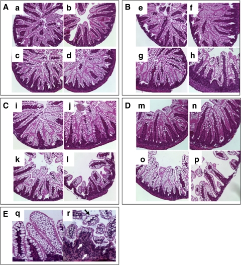
Effect of formula feeding and hypoxia on intestinal morphology. A: intestinal morphology at 24 h (n = 24, 6 rats/group). a: Dam fed. b: Dam fed-hypoxic. c: Formula fed. d: Formula fed-hypoxic. B: intestinal morphology at 48 h (n = 24, 6 rats/group). e: Dam fed. f: Dam fed-hypoxic. g: Formula fed. h: Formula fed-hypoxic. C: intestinal histological morphology at 72 h (n = 91). i: Dam fed (n = 23). j: Dam fed-hypoxic (n = 22). k: formula fed (n = 21). l: Formula fed-hypoxic (n = 25). D: intestinal morphology at 120 h (n = 24, 6 rats/group). m: Dam fed. n: Dam fed-hypoxic. o: Formula fed. p: Formula-fed-hypoxic. E: q and r with high-power image (×200). q: Vacuolated villus cells. r: Necrotizing enterocolitis (NEC) histology with villus core separation (black arrow) and infiltrating inflammatory cells (white arrows). Intestinal segments from dam-fed and dam-fed-hypoxic animals at all time points show normal histology. The villi are tall and healthy. A, B, C, and D are with magnification = ×100. Intestinal segments from formula-fed animals without hypoxic insult show mild changes (score = 1) with shorter and vacuolated villi after 72 h (k, o, and q). Intestinal segments from formula-fed rats subjected to hypoxia displayed NEC histological abnormalities (score ≥ 2) (l, p, and r) after 72 h. The median score was 2.0, with the incidence of NEC 56% (14/25) at 72 h and 50% (3/6) at 120 h.

