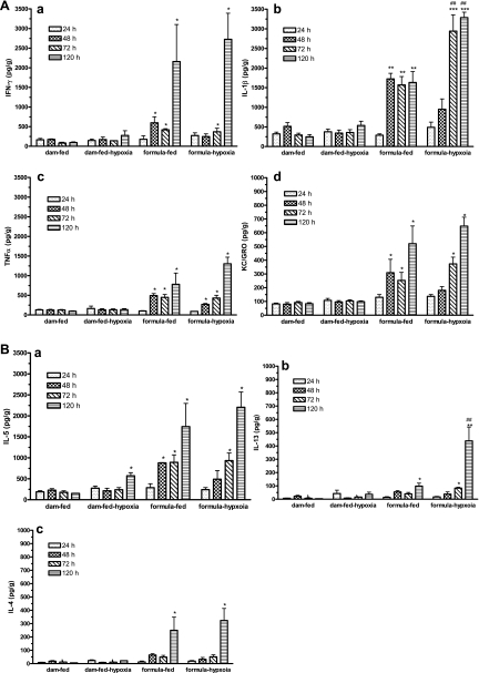
Fig. 6.
Cytokine production by normal, hypoxic, formula-fed, and NEC ileal tissues. Cytokines in ileal tissue lysates at 24, 48, 72, and 120 h exposure to formula feeding and/or hypoxia were assayed by using a MSD rat 7-spot plate (see materials and methods) to compare with dam-fed controls. Each well of the 96-well plate contained antibodies to IL-1β, IFN-γ, KC/GRO (Cxcl1), TNF-α, IL-5, IL-13, and IL-4. A: Th1 cytokines IFN-γ (a), IL-1β (b), TNF-α (c), and KC/GRO (d). B: Th2 cytokines IL-5 (a), IL-13 (b), and IL-4 (c). Data are represented as means ± SE. N = 3. All groups compared with dam-fed control: *P < 0.05; **P < 0.01; ***P < 0.001. Formula-fed-hypoxic group compared with formula-fed group: #P < 0.05, ##P < 0.01. Th1 and Th2 cytokines were produced in the intestines of both formula-fed and formula-fed-hypoxic rats at 48 h. Note that IL-1β (Th1) and IL-13 (Th2) were significantly increased in NEC compared with formula-fed controls.

