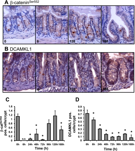
Fig. 8.
Dox treatment induces changes in the presence of β-cateninSer552 and DCAMKL1 expressing cells. Immunohistochemistry was used to identify β-cateninSer552 or DCAMKL1-positive cells 0, 6, 24, 48, 72, 96, 120, and 168 h after Dox treatment (n = 3). A: β-cateninSer552 staining at 0, 6, 120, and 168 h revealed changes in the number of positive cells that are expressed in graphic form in C. DCAMKL1 staining (B) at 0, 6, 120, and 168 h revealed changes in the number of positive cells as well, which are quantified in D. *Statistical significance compared with zero-time control (P < 0.05).

