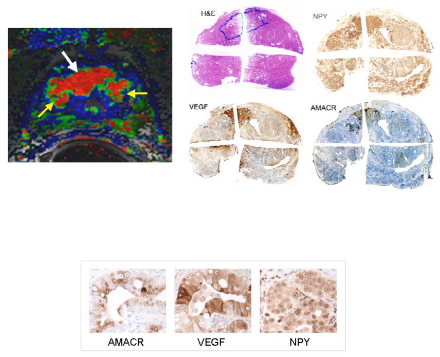
Figure 5.
Immunohistochemical stains obtained using horseradish peroxidase readout of the prostate of a patient with proven prostate cancer. The tumor is outlined in blue of the H and E stain in the series of low magnification images showing an entire slice of the gland. The color-coded image obtained from an analysis of the non-invasive DCEMRI sequence is shown on the left. The red region indicated by the white arrow is the tumor. The yellow arrows are showing two BPH nodules adjacent to the tumor. The intensity of VEGF and NPY expression shown in the high magnification images reflects the same conditions of IHC staining as shown in the high magnification images in Figure 3.

