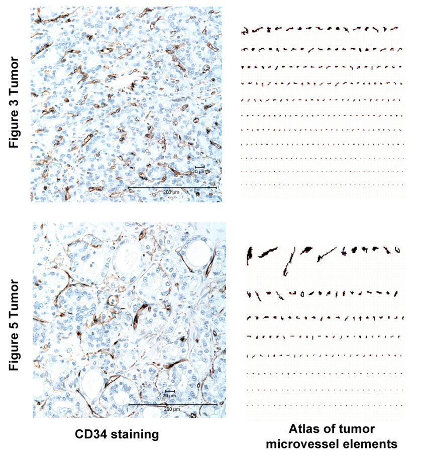
Figure 6.
Contrasting Patterns of Tumor Microvasculature in the Prostate Cancers Shown in Figure 3 and Figure 5. CD34 IHC staining was used to visualize tumor microvessels. An image analysis “atlas” of the microvessel elements highlight the difference between these two cancers, with the more uniform pattern evident in the Figure 3 tumor being characteristic of a more “mature” tumor vasculature.

