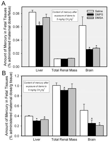
Figure 5.
Amount of mercury in organs of fetuses extracted from pregnant Wistar rats exposed to 5 mmol/kg CH3HgCl on day 17 of pregnancy and treated 24 h later with saline (2 mL/kg), DMPS or DMSA (200 mg/kg). Panel A shows the disposition of mercuric ions in organs of each fetus (% administered maternal dose per fetus). Panel B shows the disposition of mercury when factored by the weight of each organ (% administered maternal dose per gram tissue). Fetuses were harvested 48 h (embryonic day 19) after exposure of dams to CH3HgCl. Data represent mean ± SE of 11-18 fetuses and placentas from five rats. *, significantly different (p < 0.05) from the corresponding mean for rats treated with saline.

