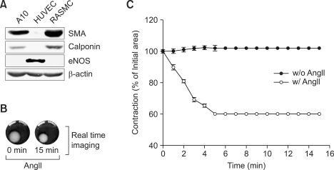
Figure 1.
AngII-induced RASMC contraction. (A) Cells from rat aortic smooth muscle were subjected to western blot analysis using the indicated antibodies. A10 and HUVEC cell lysates were included as markers for smooth muscle and endothelial cells, respectively. (B) Isolated RASMCs were embedded in collagen gel matrix as described in Methods. Gel contraction was initiated by addition of AngII (1 µM), and images were captured as described in Methods. Images are representative of two independent experiments each done in triplicate. (C) RASMC-embedded collagen gel was stimulated with either vehicle or AngII (1 µM), images were taken, and the surface area of the collagen gel was analyzed as described in Methods. Data are mean ± S.D. of two independent experiments (n = 3 for each experiment).

