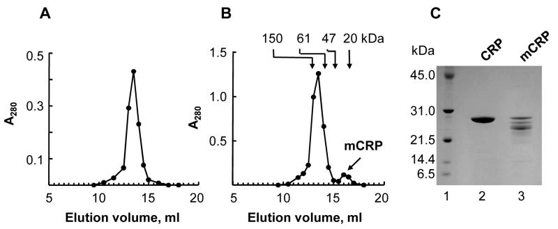
Fig. 2.
Purification of mCRP. (A) Gel filtration chromatography of freshly-purified CRP. A representative of 5 chromatograms from a Superose12 column is shown. (B) Gel filtration chromatography of stored CRP. A representative of 5 chromatograms from a Superose12 column is shown. The arrows point to the elution volumes of molecular weight marker proteins. The first peak contained CRP while the second peak contained mCRP. (C) CRP and mCRP were subjected to 10–20% SDS-PAGE under reducing conditions. A representative Coomassie blue-stained gel is shown. Lane 1, molecular weight marker; Lane 2, freshly-purified CRP (10 μg); Lane 3, purified mCRP (10 μg).

