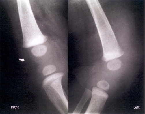
Figure 1.
Plain radiograph in lateral projection of both knee joints. Indistinctness of soft tissue-fat iterface at the left knee joint is demonstrated suggesting presence of oedema. Note the normal right side with clear fat plane (arrow) imaging. There is also a radiolucent area at the left tibial epiphysis suggesting osteomyelitis.

