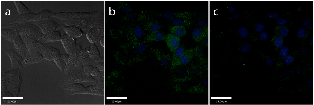
Figure 4.
Live cell images of Min6 cells after incubating with ZP3B (green) and Hoescht 33258 nuclear stain (blue) for 3 h. DIC image (a) and merged green and blue channels before (b) and after (c) the addition of TPEN. A false color image that better reveals the difference between the images in (b) and (c) may be found in Supporting Information.

