
Figure 2.
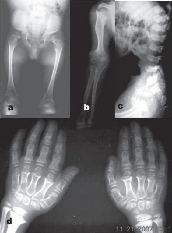
8 year old (elder) sibling. (a) Radiograph (anteroposterior view) of pelvis reveals squared ilium, narrow sacrosciatic notches, dysplastic acetabuli, and a characteristic medial beak at femoral neck. (b) Radiograph (anteroposterior view) of upper limb showing markedly flared and irregular metaphysis with deformed, irregular, and fragmented epiphyses. (c) Lateral radiograph of Lumbosacral spine showing platyspondyly with central beaking. (d) Radiograph (anteroposterior view) of hand shows underdeveloped carpals with short and broad metacarpals and phalanges