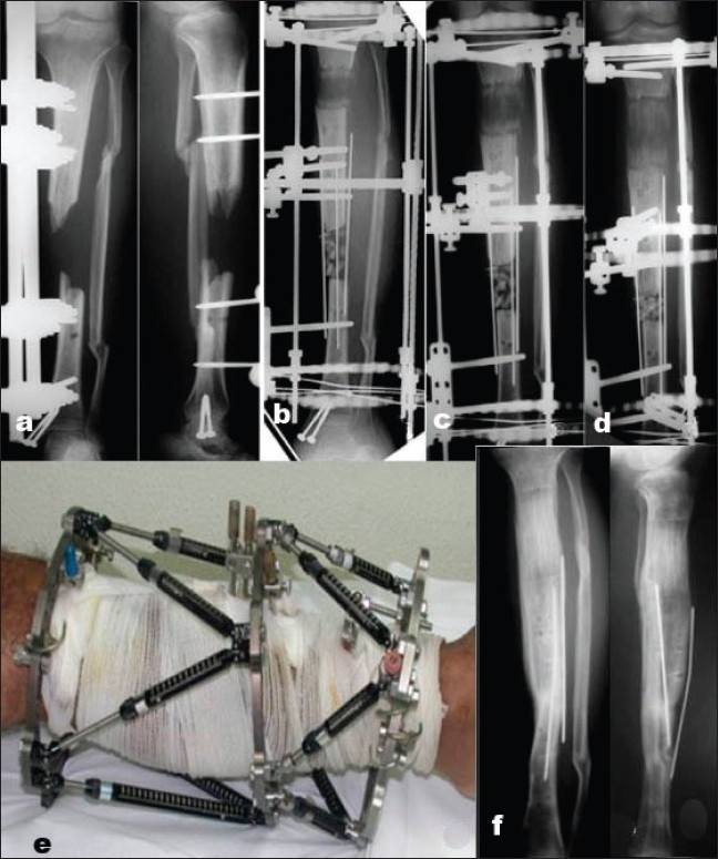
Figure 3.

(a) Anteroposterior radiographs of open tibial shaft fracture with bone loss (case 14). (b,c,d) Anteroposterior radiographs after application of the TSF and progress of bone transport. (e) Clinical picture with TSF rings and Struts. (f) Anteroposterior and lateral radiographs 3 months after fixator removal with consolidated bone transport, healed docking site, and anatomic alignment is present.
