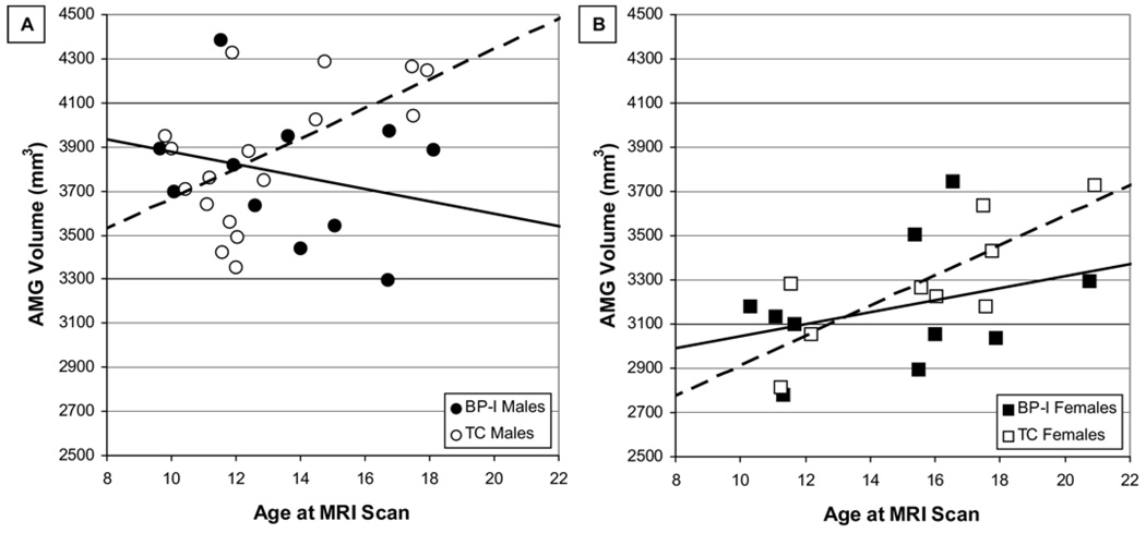
Figure 2.
Amygdala (AMG) volume by age and sex in bipolar I disorder (BP-I) and typically developing control (TC) subjects. (A) The solid line illustrates the relationship between age and AMG volume in male BP-I subjects, whereas the dashed line illustrates the relationship between age and AMG in male TC subjects. Significant predictors in a general linear model of AMG volume in male subjects were group (p = .046) and the age × group interaction (p = .028). (B) The solid line illustrates the relationship between age and AMG volume in female BP-I subjects, whereas the dashed line illustrates the relationship between age and AMG in female TC subjects. Age was a significant predictor in a general linear model of AMG volume in female subjects (p = .016). MRI, magnetic resonance imaging.

