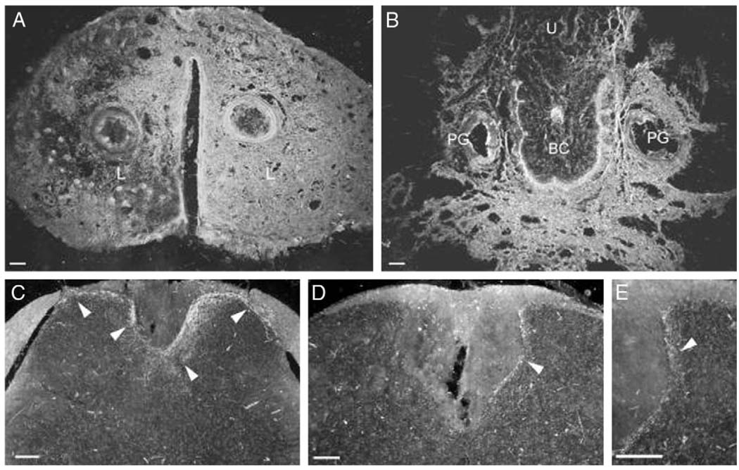
FIG. 3.
Dark field digital micrographs show labeling following large injection volume of 3 µl WGA-HRP into clitoris of 1 mouse. WGA-HRP was present bilaterally in external epithelium and clitoral sheath, that is labia (L) (A), ventral clitoris and tissue surrounding preputial glands and urethra (U), vaginal canal and gastrointestinal tract (B). Transverse sections through L2 (C) and L6 (D and E) spinal cord segments reveal resultant afferent labeling (arrowheads) of medial and lateral dorsal horns, and dorsal gray commissure. Scale bars represent 100 µm. BC, clitoral body.

