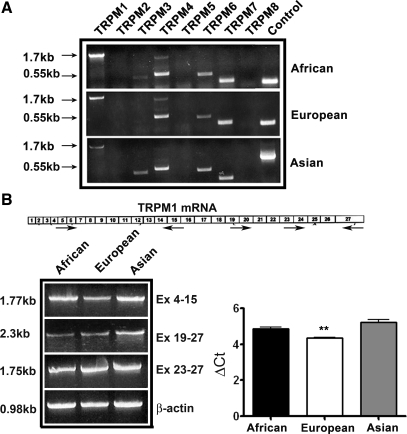
Fig. 1.
Expression of transient receptor potential melastatins (TRPMs) in human melanocytes. A: RT-PCR analysis of TRPM expression in primary melanocytes isolated from neonates of African, Asian (Korean), and European ancestry. PCR amplification of house keeping genes β-actin or GAPDH is shown in control lane. B: exon organization of TRPM1 (top) and RT-PCR analysis (bottom) of TRPM1 expression in melanocytes using primers spanning 5′ (Ex. 4–15) and 3′ ends (Ex. 19–27 and Ex. 23–27). Quantitation of TRPM1 mRNA by real-time PCR (bottom right). Data shown are ΔCt for African, European, and Asian melanocytes. **P < 0.05, Student's t-test.

