Fig. 3.
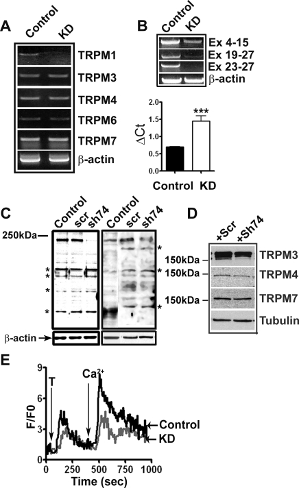
TRPM1 knockdown by short hairpin RNA (shRNA) lentivirus. A: specificity of TRPM1 knockdown was analyzed by RT-PCR using RNA isolated from cells infected with either a scrambled (control) or TRPM1 shRNA lentivirus (KD) for 48 h. B: confirmation of TRPM1 knockdown using primers spanning different TRPM1 exons (top) and real-time PCR for TRPM1 mRNA expression in control and TRPM1 knockdown cells (bottom, P < 0.05). C: knockdown of TRPM1 protein expression by shRNA. Left: HEK293 cells transfected with TRPM1-L expression plasmid infected with either a scrambled (scr) shRNA or TRPM1shRNA (sh74) lentiviruses, control without any transduction, analyzed by Western blotting with anti-TRPM1 antibodies. Right: knockdown of endogenous TRPM1 in melanocytes. Control: nontransduced; Scr: scrambled shRNA; sh74: TRPM1 shRNA. *Major nonspecific bands. D: expression of TRPMs in control and TRPM1 KD melanocytes. Numbers show the relative migration of a 150-kDa prestained molecular weight marker. E: TRPM1 knockdown decreases Ca2+ influx in neonatal foreskin melanocytes. Cells grown on glass coverslips were infected with control scrambled and TRPM1 shRNA lentiviruses loaded with Fluo-3 dye and imaged as described in materials and methods.

