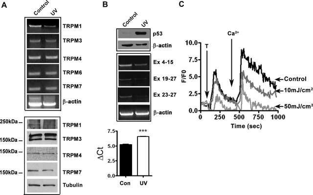
Fig. 6.
Ultraviolet B (UVB) treatment inhibits TRPM1 expression and Ca2+ influx. A: RT-PCR analysis of expression of TRPMs in control and UV-treated melanocytes (top). Effect of UV on the expression of other TRPMs analyzed by Western blotting (bottom). B: UV induced upregulation of p53 analyzed by Western blotting (top) and downregulation of TRPM1 mRNA expression (middle). β-Actin and tubulin are used as protein loading and RNA quality control, respectively. Relative quantification of TRPM1 mRNA expression in melanocytes after UV exposure (bottom ***P = 0.002) C: dose-dependent decrease in release of Ca2+ from intracellular stores and influx from extracellular medium by UVB radiation. Change in intracellular Ca2+ concentration was measured as described in materials and methods.

