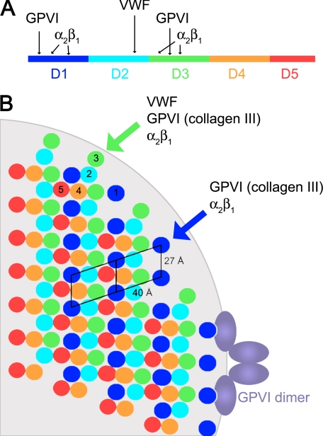
FIGURE 1.
Molecular packing of the type I collagen fiber. A, mapping the THPs specific for platelet receptors onto the collagen molecule. Each D segment is highlighted in a different color. B, schematic of the cross-section of a type I collagen fiber created by applying symmetry operators to the fiber diffraction structure (9). The cross-section was taken approximately at the beginning of segment D1. The light gray background shows the circumference of a 400-Å-diameter fiber at the same scale as the collagen triple helices. Because of interdigitation of the microfibrils, the relative position of the segments varies along the length of the fiber. The unit cell of Orgel et al. (9) was positioned such that the platelet receptor sites are accessible from the fiber surface; another possible orientation has been described (10).

