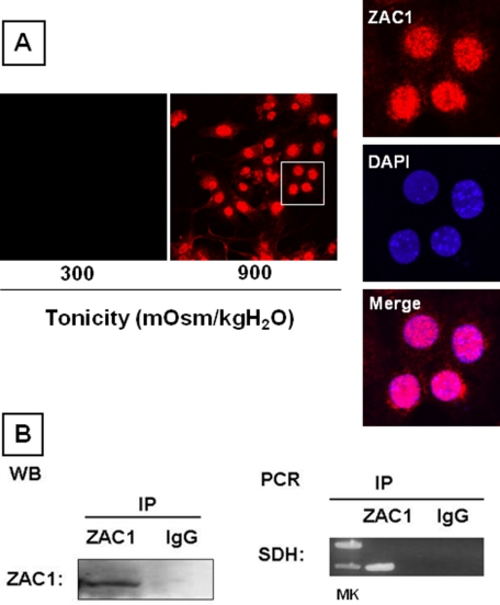
FIGURE 8.
ZAC1 represses SDH transcription. A, immunofluorescence analysis showing that ZAC1 (red) is located in the nucleus (blue) in IMCD3 cells under hypertonic conditions. DAPI, 4′,6-diamidino-2-phenylindole. B, chromatin immunoprecipitation analysis reveals that immunoprecipitated (IP) ZAC1 (left, Western blot (WB)) precipitates the repressor binding region of SDH (right, PCR) compared with the IgG control. MK, molecular mass markers.

