FIGURE 2.
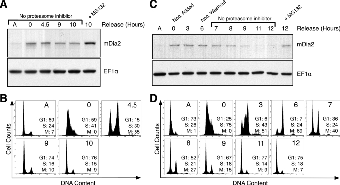
Proteasome inhibition prevents mDia2 degradation. A, HeLa cells were arrested with a double thymidine block and released into growth media. MG132 proteasome inhibitor (20 μm) was added to a population of cells upon thymidine release and incubated for 10 h (lane 6). Lysates were collected at the time points indicated and immunoblotted for mDia2. EF1α was probed as a loading control. A represents cell lysate from an asynchronous population. B, cells from A were labeled as described under “Experimental Procedures” to determine DNA content and analyzed on a flow cytometer. Plots show cell numbers relative to DNA content. The percentages of cells in G1, S, or M phase are shown for each time point. C, HeLa cells were thymidine arrested as in A (t = 0 h) but released into growth media containing 100 ng/ml nocodazole for 6 h (t = 0–6 h). Cells were then rinsed and released into normal growth media. MG132 (20 μm) was added to a population of cells 1 h after the nocodazole release and incubated for 5 h (lane 10). Immunoblotting of lysates were performed as in A. D, flow cytometry profiles showing DNA content of cells examined in C.

