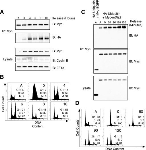
FIGURE 4.
mDia2 ubiquitination increases at the end of mitosis. A, HEK293T cells were co-transfected with Myc-mDia2 and HA-ubiquitin. Cells were arrested at G1/S phase with a double thymidine block. Cells were released into growth media, and lysates were collected at time points indicated and subjected to Myc immunoprecipitation (IP). Immunoblots (IB) were probed with Myc and HA to examine the extent of mDia2 ubiquitination. EF1α was blotted as a loading control, and cyclin E was blotted to verify progression through the cell cycle. B, cells from A were labeled as described under “Experimental Procedures” to determine DNA content and analyzed on a flow cytometer. Plots show cell numbers relative to DNA content. The percentages of cells in G1, S, or M phase are shown for each time point. C, HEK293T cells were co-transfected with Myc-mDia2 and HA-ubiquitin (second through seventh lanes) in addition to Myc-GFP and HA-ubiquitin as a negative control (first lane). Cells were incubated with 100 ng/ml nocodazole for 24 h to arrest cells in mitosis. Cells were rinsed, and lysates were collected at the indicated times after nocodazole washout. Immunoblots were probed with anti-Myc and anti-HA. D, flow cytometry profiles showing DNA content of cells from C.

