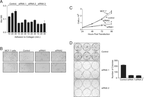
FIGURE 2.
RACK1 is required for cell adhesion, spreading, and foci formation. A, MCF-7 cells were transfected with RACK1 siRNA. After 20 h cells were serum-starved for 4 h, removed with trypsin EDTA, and resuspended in serum-free medium at 2 × 10⁵ cells/ml for analysis of adhesion to collagen-coated plates. At the indicated times cells were stained with crystal violet, and absorbance was determined at 595 nm. Data are presented for quadruplicate samples. B, MCF-7 cells were transfected with siRNA, cultured for 20 h, removed with trypsin EDTA, seeded into collagen-coated wells of a 24-well plate, and allowed to adhere for 8 h before being examined and photographed with an inverted microscope (20× magnification). C, MCF-7 cells transfected with siRNA were seeded in multiple wells of a 24-well plate at a density of 3 × 10⁴ cells/well 24 h post-transfection. At the indicated time points cells were collected, and the number and viability were determined using a hemocytometer and trypan blue exclusion. Data are presented as the mean and S.D. of live cell numbers in triplicate wells. D, MCF-7 cells transfected with siRNA were seeded in multiple wells of a six-well plate. After 24 h cells were seeded at a density of 500 cells per well in medium supplemented with 10% fetal calf serum and incubated for 14 days at 37 °C. After 14 days the cells were stained with Giemsa. The plates were photographed, and the colonies were counted. Data are presented as the mean and S.D. of colonies per well for six wells and are representative of three independent experiments with similar results.

