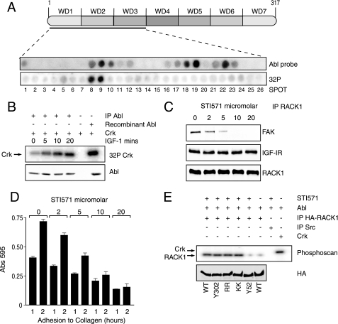
FIGURE 6.
c-Abl phosphorylates RACK1 at the FAK interaction site and c-Abl kinase activity is required for interaction of FAK with RACK1. A, A RACK1 peptide array was probed with recombinant active c-Abl, which was detected by immunoblotting with anti-c-Abl antibody. Blank spots indicate no binding and were evident in all sections of the array except for spots in WD2 (corresponding to peptides 8, 9, and 10) and WD3 (corresponding to peptides 18, 19, 22, and 23) (top). To measure phosphorylation of the RACK1 array by c-Abl kinase, the membrane was incubated in 20 ml of membrane phosphate buffer together with 1 μg of c-Abl and 32P at 30 °C for 30 min. After extensive washing, radioactivity was detected with phosphorimaging. The spots were blank in all sections of the array except for spots corresponding to peptides 8 and 9 in WD2. B, c-Abl was immunoprecipitated (IP) from MCF-7 cells which had been serum-starved for 4 h and stimulated with IGF-I for the indicated times. The immunoprecipitated proteins were subjected to an in vitro kinase assay using GST-Crk as a substrate. The total amount of c-Abl used in the kinase assay was detected by Western blotting. C, MCF-7 cells maintained in serum were pretreated with increasing concentrations of STI571. RACK1 was immunoprecipitated (IP) from the lysates and investigated for associated FAK before probing with antibodies against the IGF-IR and RACK1 to show that equal amounts of the protein were immunoprecipitated from each sample. D, MCF-7 cells maintained in serum were pretreated with increasing concentrations of STI571. The cells were then serum-starved for 4 h and resuspended in serum-free medium and allowed to adhere to collagen coated plates for the indicated times before staining with crystal violet and analysis of absorbance at 595 nm. Data are presented for quadruplicate samples. E, MCF-7 cells were transfected with plasmids encoding HA-RACK1 WT, Y302F, R57A/R60A, K127A/K130A, or Y52F. Cell lysates were prepared and immunoprecipitated with anti-HA antibody and used as a substrate in an in vitro kinase assay with c-Abl kinase. Recombinant Crk and immunoprecipitated Src were used as positive and negative controls, respectively. Incorporated radioactivity was detected by phosphorimaging.

