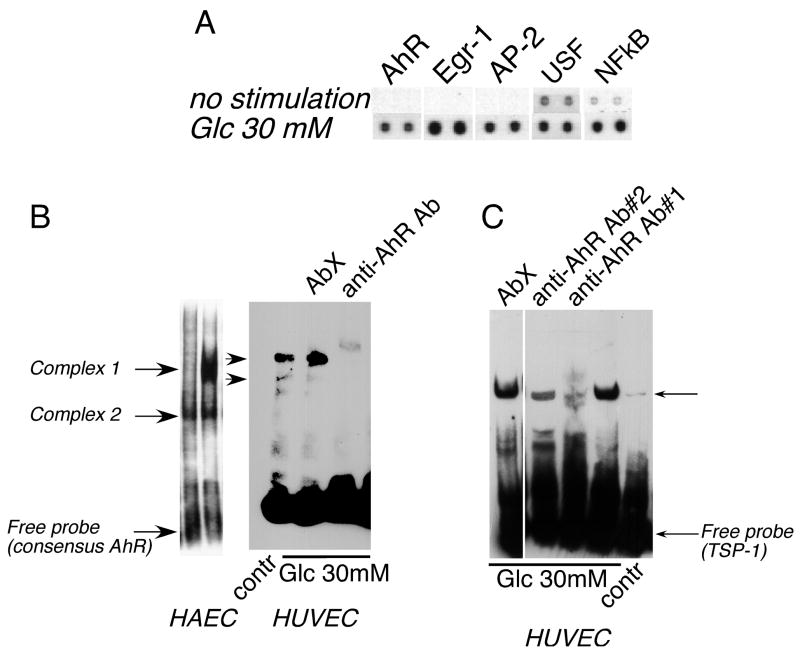
Figure 2. AhR is activated in endothelial cells in response to high glucose.
A Nuclear extracts from HAEC stimulated with glucose for 1 hour were analyzed in a Protein/DNA array (Panomics) to detect activated transcription factors. Representative results for AhR, Egr-1, AP-2, USF-1 and NFkB (transcription factors with putative binding sites in the −280/+66 fragment of the THBS1 promoter) are shown. B: Activation of AhR was confirmed in EMSA (5μg of nuclear extract) using the consensus AhR probe. Formation of complexes was prevented by anti-AhR antibody RPT1 (1 μg) but not an unrelated antibody (AbX). C: The predicted binding site for AhR in the promoter fragment responsive to glucose (−280/+66) was confirmed in EMSA using anti-AhR antibody.

