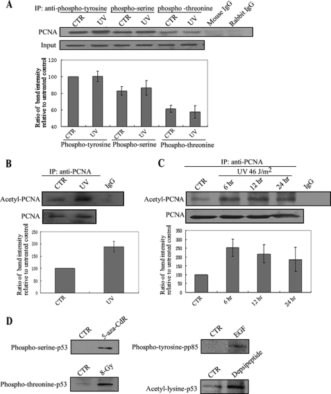
FIGURE 4.
UV irradiation induces PCNA acetylation in A549 cells. A, A549 cells were irradiated with UV at 46 J/m2 and incubated for 6 h. Protein was extracted and then immunoprecipitated using anti-phosphotyrosine, phosphoserine, or phosphothreonine, followed by Western blotting using anti-PCNA. Mouse or rabbit IgG was used as a negative control. PCNA bands were scanned, and relative band intensities were normalized for each input band. The lower column diagram represents the average relative band intensity of PCNA with standard error from several independent experiments. B and C, A549 cells were irradiated with UV at 46 J/m2 and incubated for 6 h (B), or at 23 J/m2 for different incubation times (6, 12, or 24 h) (C). Protein was extracted and then immunoprecipitated using anti-PCNA followed by Western blotting using anti-acetyl-lysine. IgG was used as a negative control. The representative value of the lower column diagram is the average relative band intensity of PCNA with standard error from several independent experiments. D, epidermal growth factor (EGF) was added to human A431 carcinoma cells to identify phosphotyrosine in pp85. A549 cells were treated with 5-aza-CdR (0.1 μm, 48 h), x-ray (8-Gy), or depsipeptide (0.1 μm, 6 h) to test for phosphoserine, phosphothreonine, or acetyl-lysine of p53, respectively.

