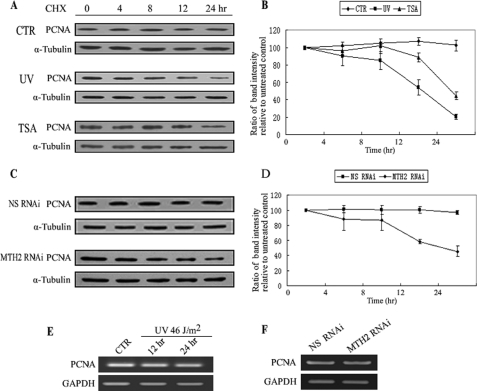
FIGURE 6.
Half-life of PCNA is shortened after dissociation of PCNA and MTH2. A and B, CHX was added into untreated, UV-treated, or TSA-treated A549 cells at 25 μg/ml for different times as indicated. Western blotting was performed from whole cell extracts with anti-PCNA (A). The hyphenated line graph shows the change in PCNA level at different times. The representative value is the average band intensity of PCNA with standard error from several independent experiments (B). C and D, 24 h after NS-siRNA or MTH2-siRNA was transfected into A549 cells, CHX was added to the medium at 25 μg/ml for various lengths of time as indicated. NS-siRNA was used as negative control for the RNAi assay. The hyphenated line graph shows the change in PCNA level at different times. The representative value is the average band intensity of PCNA with standard error from several independent experiments (D). E and F, A549 cells were irradiated with UV at 46 J/m2 and then incubated for 12 h or 24 h (F). NS-siRNA or MTH2-siRNA was transfected into A549 cells for 48 h (G). mRNA was extracted for RT-PCR.

