FIGURE 2.
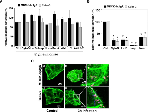
Invasion of pneumococci via pIgR requires the dynamics of the host cell actin cytoskeleton and microtubuli. A, pneumococcal adherence to MDCK-hpIgR and Calu-3 cells was determined in the absence (control, Ctrl) or presence of various inhibitors. Adherence of S. pneumoniae in the absence of an inhibitor was set to 100%. B, invasion of MDCK-hpIgR or Calu-3 cells with pneumococci was followed in the absence (control) or presence of inhibitors of actin filaments and microtubules, including cytochalasin D (CytoD, 125 nm), latrunculin B (LatB, 50 nm), jasplakinolide (Jasp, 100 nm), and nocodazole (Noco, 10 μm), by the antibiotic protection assay. Invasion of S. pneumoniae in the absence of an inhibitor was set to 100%. *, p < 0.001 relative to infections carried out in absence of an inhibitor. C, immunofluorescence microscopy illustrating changes of the actin cytoskeleton after infecting host cells for 3 h with pneumococci. F-actin was stained with AlexaFluor-488 phalloidin, and intracellular pneumococci were stained with AlexaFluor-568 (red), whereas adherent bacteria were stained with Cy5 and hence, bacteria appear pink (blue/red stain). Uninfected host cells (a and d) and infected host cells (b and e), respectively. Higher magnifications (c and e) illustrate changes of the actin cytoskeleton during pIgR mediated infection of MDCK-hpIgR or Calu-3 cells.

