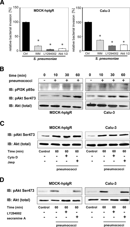
FIGURE 6.
PI3K and Akt are activated during pneumococcal invasion via pIgR. A, invasion and intracellular survival of the bacteria in MDCK-hpIgR and Calu-3 cells were monitored in the absence (Ctrl) or presence of PI3K inhibitors wortmannin (50 nm) or LY294002 (50 μm) and in the presence of Akt Inhibitor VIII (Akti, 10 μm) by the antibiotic protection assay. Pneumococcal invasion in the absence of the inhibitor was set to 100%. *, p < 0.001 relative to infections carried out in the absence of inhibitor. B, pneumococcal infection of MDCK-hpIgR and Calu-3 cells induces phosphorylation of PI3K subunit p85α and Akt. Host cell lysates of MDCK-hpIgR and Calu-3 cells prepared after pneumococcal infections and separated by 10% SDS-PAGE. Activation of kinases were analyzed using antibodies against phosphorylated forms of PI3K subunit p85α (upper panel) or Akt (pAkt) (middle panel). The membrane was stripped and reprobed with total Akt antibody and used as loading control (lower panel). C, activation of Akt is independent of pneumococcal invasion. Pneumococcal invasion was blocked by inhibition of the actin cytoskeleton dynamics with Cytochalasin D (CytoD) or jasplakinolide (Jasp) and activation of Akt followed. Total Akt served as loading control (lower panel). D, Akt is activated in a PI3K-dependent but Cdc42-independent manner. Activation of Akt was followed in the presence of PI3K inhibitor LY294002 (50 μm) or Cdc42 inhibitor secramine A (10 μm). Total Akt served as loading control (lower panel).

