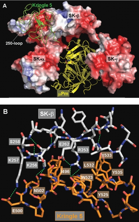
FIGURE 4.
Putative model of plasminogen K5 recognition by SK β-domain. A, overall assembly of the ternary K5·SK·μPm complex predicted by FTDOCK. The SK moiety is shown as a solid surface model colored according to its electrostatic surface potential (blue, positive; red, negative), and the bound μPm is represented as a yellow ribbon. SK domains are indicated. The kringle domain of the incoming Pg substrate is given as a green ribbon. B, close-up of interactions between SK 250-loop and human Pg kringle 5 module. For simplicity, only residues of SK 250-loop and neighboring K5 residues are included in this plot. Hydrogen bonds are shown as green dotted lines. Molecular modeling was performed as described under ”Experimental Procedures.“

