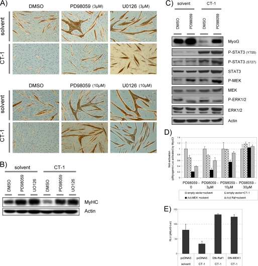
FIGURE 5.
CT-1 inhibits the transcriptional activity of the MRFs through activation of MEK signaling. A, C2C12 cells were plated at equal density and induced differentiation transferred into DM upon about reaching confluence. The cells were maintained in indicated concentration of MEK inhibitor (PD98059, U0126, or DMSO; 3 μm or 10 μm) with without CT-1 (10 ng/ml). After 2days in the indicated conditions, the cells were fixed and stained for MyHC detection by immunochemistry with MF-20 mouse monoclonal antibody. MyHC protein accumulation was indicated by brown color. The photomicrographs are representative fields. B and C, C2C12 cells were maintained in DM with CT-1 (10 ng/ml) and or PD98059 (10 μm), or their solvents for 2days (C) or 3days (B) to induce myotube formation. Total cellular proteins were extracted from the cells in each condition. The total protein lysate samples (20 μg) were subjected to Western blotting analysis. Actin levels indicate loading of an equal amount of the total protein into each lane. D, C2C12 cells were transfected with a pMyoG-Luc (0.5 μg), a MyoD expression vector (1 μg), a pCMV-β-Gal (0.3 μg), and also the indicated kinase expression vector (act.MEK1, act.Raf) or an empty vector (1 μg). The transfected cells were maintained in DM containing CT-1 (10 ng/ml) or solvent, and the indicated concentration of PD98059 MEK inhibitors for 16 h. The cells were harvested and subjected to Luciferase assay and β-Gal assay. Luciferase activity was normalized according to the β-galactosidase activity from a co-transfected pCMV-β-Gal expression construct by calculating the Relative Luciferase Unit (relative luciferase unit) for each individual condition, and the fold-activation was calculated with respect to the average relative luciferase unit of the “empty vector + solvent” at the corresponding concentration of PD98059. E, C2C12 cells were transfected with a pMyoG-Luc (0.5 μg), a pCMV-β-Gal (0.3 μg), and also the indicated kinase expression vector (DN-MEK1, DN-Raf1) or an empty vector (1 μg). The transfected cells were maintained in DM containing CT-1 (10 ng/ml) or solvent for 16 h. The cells were harvested and subjected to Luciferase assay and β-Gal assay.

