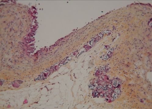
Figure 13).
World Health Organization stain section of the capsule around the injected polyacrylamide hydrogel material. The interface surface of the capsule (at top of image) is covered by a row of mono-nuclear and multinucleated histiocytes. These cells are supported by multiple layers of collagenous fibrous tissue. On the surface of the capsule and within the capsule, there are pools of amorphous, nonbirefringent, granular, foreign material (original magnification × 12.5)

