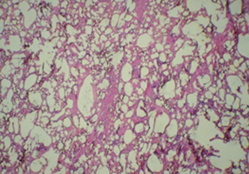
Figure 5).
Histology of hematoxylin and eosin staining of the tissue shown in Figure 4. There is extensive involvement of the breast tissue by silicone, which appears as empty spaces or vacuoles filled with silicone. There are occasional multinucleated giant cells, areas of vascular obliterans, chronic inflammation, and destruction of breast parenchyma (original magnification ×50)

