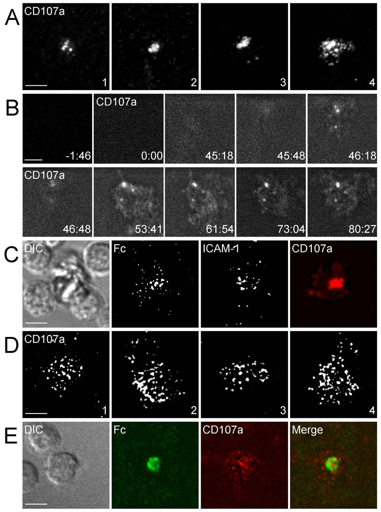
Figure 3. Central Accumulation of LAMP-1 in Synapses Formed over Ligands for LFA-1 and CD16.
Degranulation was monitored by TIRFM with a soluble CD107a F(ab) conjugated with Alexa Fluor 647. Scale bars are 5.0 µm. The images are representative of at least 50 cells in five independent experiments. (A and B) Live NK cells imaged on bilayers carrying unlabeled ICAM-1 and IgG1 Fc. (A) Individual cells imaged at ~50 min (#1), ~80 min (#2), ~120 min (#3), and ~26 min (#4), after addition to the bilayer. (B) Time series taken from Movie S8. The first frame was taken prior to the addition of CD107a Fab and of NK cells. The second frame was taken at the time of CD107a Fab and NK cell injection into the chamber. (C) NK cells imaged at ~120 min after addition to a bilayer carrying Alexa Fluro 488 conjugated ICAM-1 and Alexa Fluor 568 conjugated Fc. (D and E) Live NK cells imaged on bilayers carrying IgG1 Fc alone. (D) Individual cells imaged at ~30 min (#1), ~40 min (#2), ~60 min (#3), and ~100 min (#4) after addition to the bilayer. (E) NK cells imaged at ~30 min after addition to a bilayer carrying Alexa Fluor 488-conjugated Fc.

