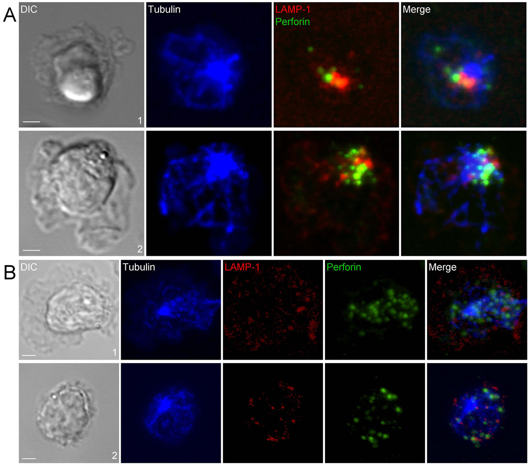
Figure 7. Polarized Perforin-Containing Lytic Granules Contact Internalized LAMP-1 in the Presence of ICAM-1.
(A) NK cells imaged on bilayers containing Fc and ICAM-1 ~60 min (#1) and ~120 min (#2) after injection over the bilayer. Perforin (Green), internalized LAMP-1 (Red), and Tubulin (Blue) were acquired by 3D confocal microscopy. Scale bars are 5.0 µm. The images are representative of at least 30 cells in two independent experiments. (B) NK cells imaged on bilayers carrying Fc ~120 min after injection over bilayer. Differential interference contrast (DIC) images are shown on the Left. Merged overlays of fluorescent are on the Right. LAMP-1(Red) detected by inclusion of 0.016 µM Alexa Fluor 647-labeled CD107a Fab during NK cell incubation on with lipid bilayer containing Fc, MTOC (Blue) and perforin (Red) are shown in Center. Scale bars are 2.0 µm. The images are representative of 50 cells in three independent experiments.

