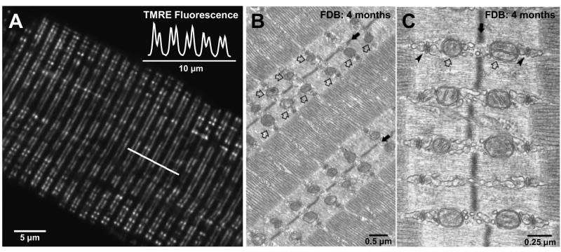
Figure 1. Mitochondria are precisely localized adjacent to the calcium release unit in adult mammalian fast-twitch skeletal muscle.
A. Confocal image of a single flexor digitorum brevis (FDB) skeletal muscle fiber obtained from an adult mouse (4 months of age) stained with the mitochondrial-selective dye tetramethylrhodamine ethyl ester (TMRE). TMRE fluorescence along the line of interest marked in A shows characteristic doublets of fluorescence with a sarcomeric periodicity of ~2 μm (inset). B. and C. Representative low (B) and high (C) resolution electron micrographs of FDB skeletal muscle fibers obtained from an adult mouse (4 months of age). Mitochondria (open arrows) are aligned adjacent to the triad (arrowheads), on either size of the Z-line (solid arrows). Images kindly provided by Drs. Simona Boncompagni and Feliciano Protasi.

