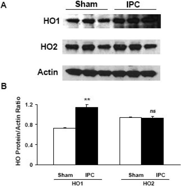
Fig. 3.
Ischemic preconditioning (IPC) increased HO1 protein expression levels after 24 h in the brain cortex of wildtype mice. (A) Western blots were performed to measure the protein expression of HO1 and HO2. The expression of actin was used as a loading control. HO2, which is known to be constitutively expressed in the brain, was used as a control and, as expected, was unaffected. (B) The histograms show the ratio of density captured from HO1 and HO2 to that of actin. Values shown are means ± SEM from three independent sets of experiments. **p < 0.01 vs. corresponding control; ns, not significant.

