
Figure 1.
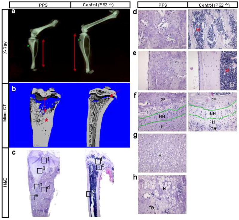
Skeletal phenotype of PPS mice at 8 weeks of age. (a) X-ray radiographs of hindlimbs. Red double-headed arrows denote length of tibia. Green arrowheads point to trabecular bone region. (b) Medial, longitudinal section through 3-D reconstruction of the tibia by μCT. Double-headed arrow: expanded growth plate; asterisk: excessive bone; arrow: normal growth plate. (c) H&E staining of medial longitudinal sections through the tibia. (d-h) Higher magnification of boxed areas in c. 2°: secondary ossification center; NH: nonhypertrophic region; H: hypertrophic region; TB: trabecular bone; M: marrow.