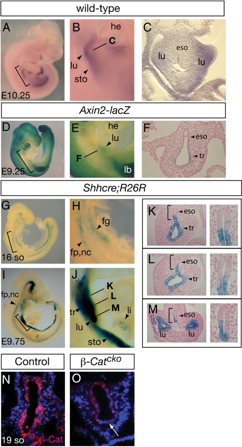
Fig. 1.
WNT/β-Catenin signaling and β-Catenin inactivation in the foregut using Shhcre. (A–C) Wnt2 expression as determined by RNA in situ hybridization in E10.25 embryos. Bracketed region in A is magnified in B, and line in B indicates the approximate level of transverse section shown in C. Axin2 is expressed in the ventral foregut mesenchyme adjacent to nascent lung buds. (D–M) β-galactosidase (β-gal) staining in Axin2-lacZ (D–F) or Shhcre;R26R embryos (G–M) at stages indicated. Bracketed regions in D, G, and I are enlarged in E, H, and J, respectively. Lines in E and J indicate approximate level of transverse sections shown in F and K–M. Bracketed regions in the left panels of K–M are magnified in the corresponding right panels. Arrowhead in E indicates Axin2-lacZ activity in the prospective respiratory region. Shhcre activity in the foregut is first detected at the 16-so stage (≈E8.75), around the time of specification. By E9.75 (after lung budding), its activity is detected in the primary lung buds, trachea, ventral esophagus, ventral stomach, intestine and isolated cells in the liver primordium. (N and O) Anti-β-Catenin antibody staining in transverse sections of 19-so stage embryos at the foregut level. Arrow in O indicates diminished β-Catenin staining in the ventral foregut epithelium of the β-Catcko mutant lung. For transverse sections shown in all figures, dorsal is up and ventral is down. Abbreviations: eso, esophagus; fg, foregut; fp, floor plate; he, heart; lb, limb bud; li, liver; lu, lung; nc, notochord; sto, stomach; tr, trachea.

