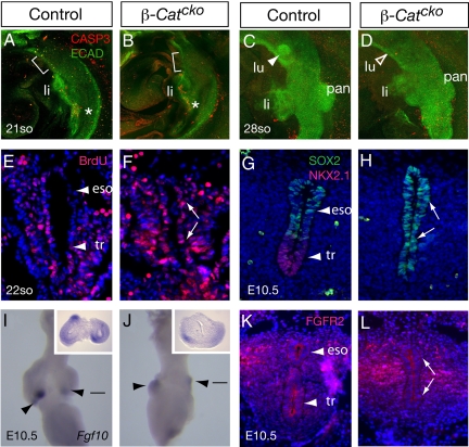
Fig. 3.
Inactivation of β-Catenin leads to a defect in foregut patterning. (A–D) Double immunofluorescence staining with anti-cleaved Caspase 3 antibody that labels cells undergoing apoptosis (red) and anti-E-Cadherin antibody that labels foregut epithelium (green) at the 21-so stage (specification) or 28-so stage (budding morpohogenesis). Lateral views are shown. Brackets in A and B indicate prospective trachea/lung region. There is no detectable increase in cell death in the mutants compared to controls. Asterisks in A and B indicate cleaved Caspase 3-positive cells just posterior to the liver in both the mutant and the control to show that the assay is working. (E and F) Assays for BrdU incorporation in transverse sections of the foregut at the 22-so stage. No difference in the percentage of positive cells is detected. (G and H) Double immunofluorescence staining with anti-NKX2.1 antibody (red) and anti-SOX2 antibody (green) in transverse sections of the common trachea/esophageal tube at E10.5. In the β-Catcko mutant, NKX2.1 expression is downregulated, and SOX2 expression is expanded to the ventral epithelium. (I and J) Fgf10 expression as detected by RNA in situ hybridization in E10.5 foregut. Ventral views are shown. Filled arrowheads indicate presence of gene expression. Lines indicate approximate level of transverse sections shown in respective insets. (K and L) FGFR2 expression as detected by an anti-FGFR2 antibody in transverse sections of trachea/esophageal region at E10.5. In the β-Catcko mutant, FGFR2 remains expressed in both dorsal and ventral foregut endoderm as indicated by arrows. Abbreviations: same as Fig. 1 with the addition of CASP3, cleaved Caspase 3; ECAD, E-Cadherin.

