Figure 6.
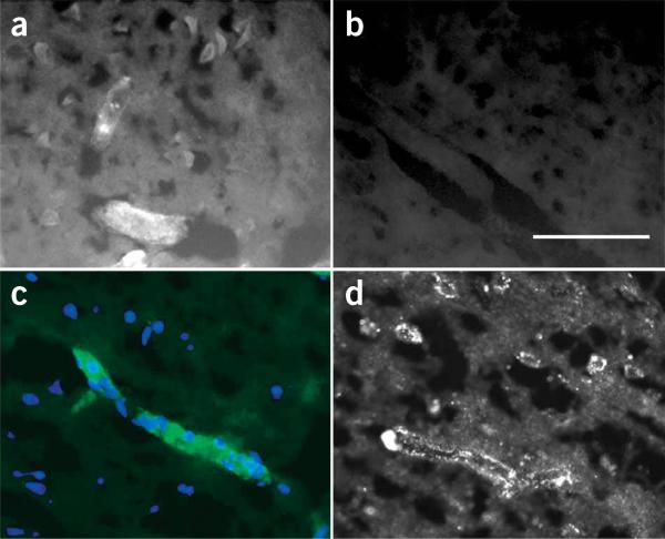
Tissue distribution of BODIPY-glibenclamide in MCA stroke model. (a–c) Fluorescence images of brain sections in a rat 8 h after MCAO (MCE model) and administration of BODIPY-glibenclamide. Fluorescent labeling was evident in cells, microvessels (a) and capillaries (c) from ischemic regions, but not in the contralateral hemisphere (b). Scale bar in b, 100 μm. Images in a,b are from the same rat, taken with the same exposure time. In c, the single layer of nuclei labeled with DAPI (blue) confirms that the structure brightly labeled by BODIPY-glibenclamide (green) is a capillary. (d) Immunofluorescence image of a brain section from a rat 8 h after MCAO (MCE model) labeled with SUR1-specific antibody showing strong labeling in a capillary and in adjacent neuron-like cells.

