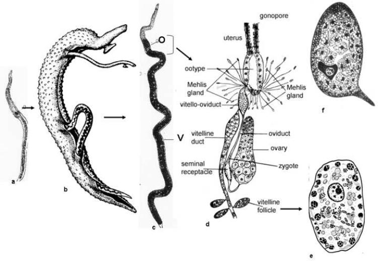
Figure 1.
Female Schistosoma mansoni, showing (a) an immature female from a single-sex infection that is stunted and reproductively underdeveloped, (b) female lying in the gynecophoric canal of a male, (c) a mature female from a bisexual infection showing that the posterior 2/3rds of the body is comprised of the vitellaria (V) that develops in response to pairing; O, ovary (d) enlargement of the mature female reproductive system. Oocytes produced in the ovary are released into an oviduct. A dilitated region in the oviduct, the seminal receptacle, is the site of fertilization. The fertilized egg moves down the oviduct to a region where it meets the vitelline duct, the vitello-oviduct. Here, each fertilized egg is surrounded by approximately 38 vitelline cells. This bolus of material moves into the ootype which is filled with mucus secretions presumably from the Mehlis' gland. With the contraction of the ootype, which determines the shape of the egg, granules are released from the surrounding vitelline cells. The materials from the granules (vitelline droplets) begin to crosslink, due to the action of a tyrosinase enzyme(s) and the eggshell forms. The egg (f) which contains the fertilized egg and a number of vitelline cells that each contain nutrients for embryogenesis is released into the uterus and one at a time deposited by the female worm through the gonopore, (e) Mature vitelline cell (S4) containing the vitelline droplets (VD) and (f) just formed egg containing the zygote and numerous vitelline cells that will contribute to embryonation. (Modified from [41])

