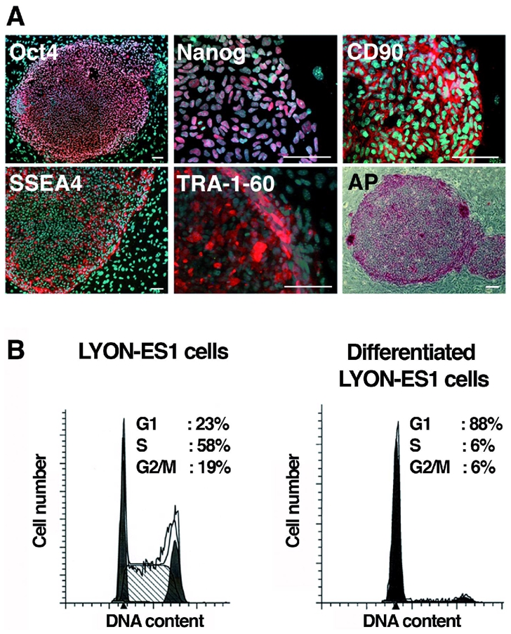
Figure 2. Characterization of the LYON-ES1 cell line.

(A) Immunofluorescent staining for Oct4, Nanog, CD90, SSEA-4, TRA-1-60, and staining for alkaline Phosphatase (AP). (B) Histograms showing cell-cycle distribution of LYON-ES1 cells and LYON-ES1 differentiated derivatives as measured by flow cytometry. Scale bars=100μm (A).
