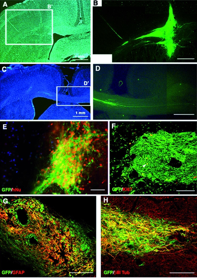
Figure 7. Integration of tau-GFP LYON-ES-derived neural precursors 28 days after transplantation in the adult rat brain.

(A, C) Low power microphotographs of hoechst stained brain sections (B) High magnification of field B′ showing the site of the grafted cells. (D) Higher magnification of the field D′, showing tau-GFP expressing axons crossing the interhemispheric border (C); (E) Colocalization of tau-GFP and human nuclear antigen (hNu). (F) Few tau-GFP cells express the proliferative marker Ki67 (yellow, arrow). Coexpression (yellow) of tau-GFP and neuronal marker MAP2 (G), and astroglial marker GFAP (H). Scale bars= 500μm (A–D); 50 μm (E–H).
